Understanding OCT Eye Exams: Advanced Retinal Imaging
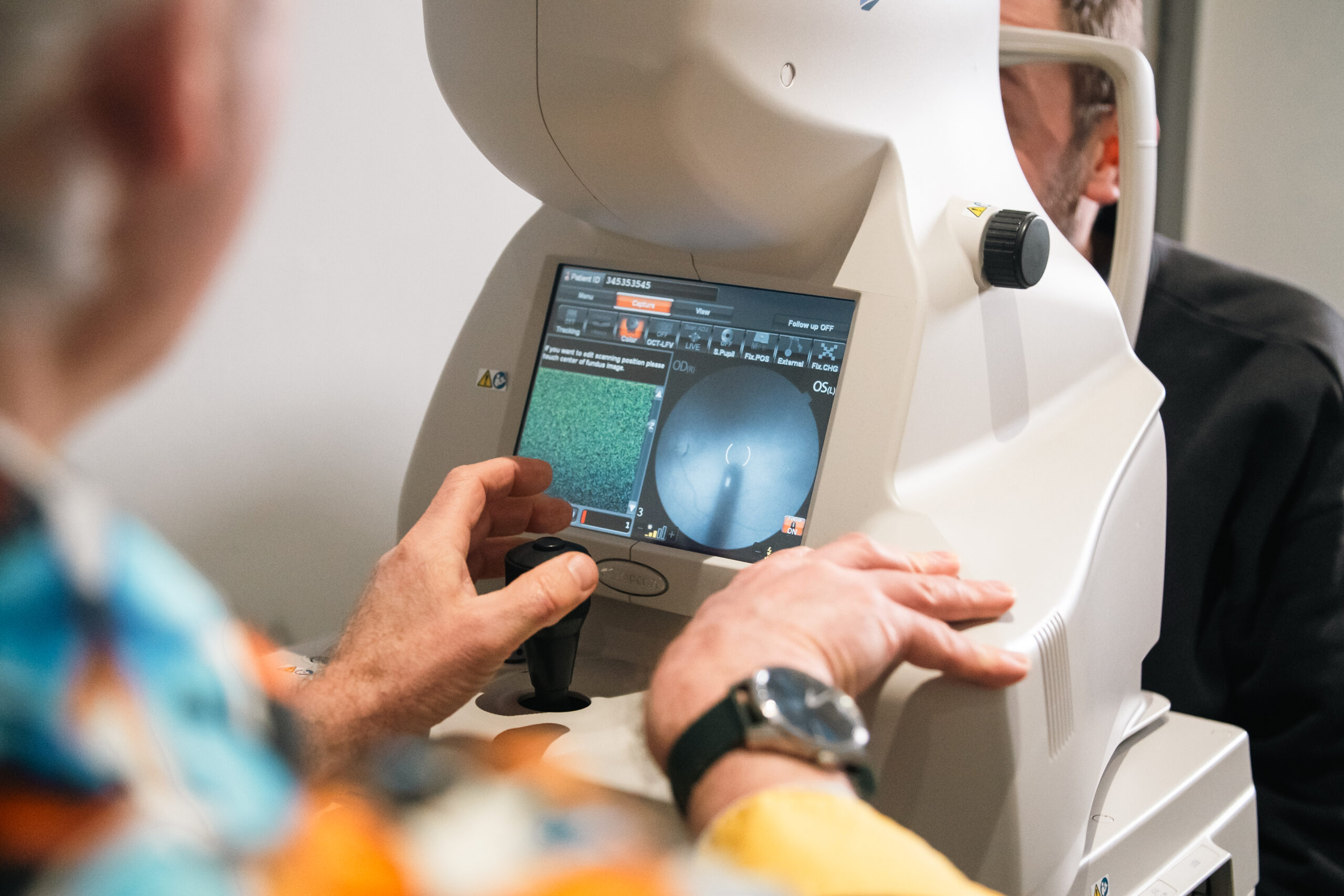
Optical Coherence Tomography (OCT) is a non-invasive, and very safe, imaging technique used in eye exams to capture detailed cross-sectional images of the retina. It is often compared to medical ultrasound because the principles are somewhat similar (one uses sound and the other uses light.) OCT is widely used in Ophthalmology to obtain cross-sectional information of the structures of the eye. It can be used to gain more information about whichever part of the eye we point it towards.
OCT is essential in assessing the health of the macula (the centre part of the eye,) but can also be used to track changes within the optic disc when there is a suspicion of glaucoma or measuring the thickness of the cornea. As a profession we are still learning about its potential and recent research indicates it may even prove to be useful in the early diagnosis of dementia or Parkinson’s disease.
We were one of the first Optometry Practice to install OCT technology. We bought our first device in 2011 and continue to be involved in new developments of the technique.